
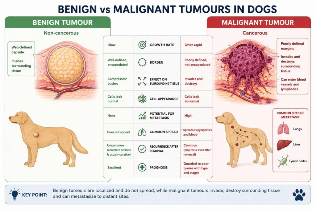
70–85% of sudden lumps on dogs under the skin are benign, however differentiating whether they are fatty deposits or abnormal cell growth requires professional diagnosis. Early diagnosis can lower the risk by up to 60% for malignant cases.
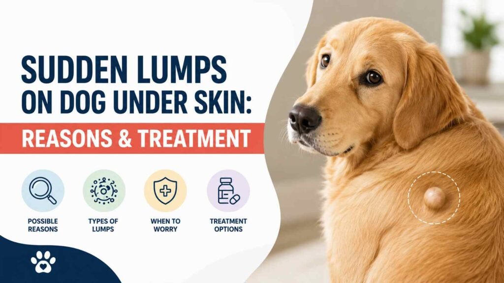
If you find a lump under your dog’s skin, it can be terrifying for a pet owner. In most cases, they are flooded with too many questions, whether it is cancer or an emergency condition, or how to treat it.
In this article, I will provide a detailed guide to these tumours, their diagnostic procedures, and treatment options, with proper research-based analysis and reviews, along with treatment costs, as reviewed by certified veterinary oncologists.
Why This Topic Matters?
According to a landmark survey by the American Animal Hospital Association (AAHA), the majority of canine lumps under the skin go undiagnosed in U.S. veterinary clinics. The “wait and see” culture is costing pets precious time when early intervention could be life-saving with proper diagnosis and treatment.
However, 2024-2025 has brought revolutionary advances in canine oncology:
- STELFONTA® is an FDA-approved intratumoral injection that is very effective against mast cell tumors without surgical intervention.
- Monoclonal antibody therapy (Gilvetmab) is widely used in cancer immunotherapy.
- Improved TKIs (Tyrosine Kinase Inhibitors) have shown good outcomes in cancer treatment; however, they can cause side effects.
- AI-Assisted Cytology is a diagnostic procedure that improves FNA accuracy from 70 to 95% to over 98%.
Statistics & Research Data
| Condition | Prevalence Among Canine Skin Masses | Typical Age of Onset | Malignancy Rate |
|---|---|---|---|
| Lipoma (Fatty Tumour) | 16% (most common benign mass) | Middle-aged to senior (7+ years) | <1% (rarely becomes liposarcoma) |
| Mast Cell Tumour (MCT) | 16-21% (most common malignant skin tumour) | Average 8-9 years; can occur younger | Varies by grade: Low (Grade I) to High (Grade III) |
| Sebaceous Gland Tumours | 7.5-12% | Older dogs (10+ years) | Mostly benign; adenomas are common |
| Histiocytoma | ~6% | Young dogs (<3 years) | Benign (regresses spontaneously) |
| Papillomas (Warts) | 5-8% | Puppies/young adults | Benign (viral origin) |
| Follicular Cysts | 4-6% | Any age | Benign |
| Abscesses | 3-5% | Any age (often after trauma/bite) | Benign (infectious) |
| Soft Tissue Sarcomas | 2-5% | Middle-aged to older | Locally invasive; metastatic potential varies |
The following are some critical survival statistics according to the tumour grade that you should know,
Despite aggressive local surgery along with systemic chemotherapy, 75% of dogs with high-grade mast cell tumors (lumps) experienced disease progression. This highlights the critical need for earlier diagnosis while tumors are still low-grade, improving the chances of effective systemic therapy.
Causes of Sudden Lumps Under Dog Skin
Mostly lumps appear under the dog’s skin when there is excess growth of cells, any fluid, foreign particles, or some other chemical substance that accumulates in a particular area of the subcutaneous space.
1. Follicular Cyst
Mostly, follicular cysts are benign in nature. These are movable, round, and may contain fluid that appears bluish through the skin. It usually occurs when hair follicles are blocked, and debris, foreign particles, or other infections can get trapped inside.
They behave like slow-growing masses that might rupture and reform over time, and are usually less painful until they become infected. They can be treated by draining the fluid; however, they often recur unless surgical intervention is made.
2. Sebaceous Cysts
Sebaceous cysts are non-movable, non-tumorous growths that are often small, round bumps with a dark centre and can produce waxy discharge. The main cause is blocked sebaceous glands, which can appear anywhere but are most common on the back of the neck or the eyelids. A small surgical CN is performed if it is enlarged or becomes infected.
3. Abscesses
An abscess can develop overnight within 24 to 48 hours. These are swollen, red, hot to the touch, and oozing pus when dogs lick the area due to pain. The main cause could be a bacterial infection from bite wounds, foreign bodies, or other ruptures.
The most effective treatment for an abscess is drainage, antibiotics, and proper wound care.
4. Seromas / Hematomas
These appear as fluid-filled swellings after surgery or trauma. These are soft and typically not painful. They occurred due to the accumulation of serum (seroma) or blood (hematoma) within the tissue space.
When the seromas or hematomas are small, they can resolve spontaneously; however, larger ones require proper veterinary drainage. They could recur unless the underlying cause his properly addressed.
5. Granulomas
Granulomas appear as firm nodular masses formed by the immune system to wall off foreign material or chronic irritation. The cause could be any foreign body, a fungal infection, or an aberrant immune response.
The only treatment is surgical removal of the granuloma, and a long-term antifungal medication could be required if the infection is confirmed.
6. Lipomas (Fatty Tumours) — MOST COMMON BENIGN MASS
Lipomas are soft, doughy, movable lumps that are usually located between the skin and muscles but not attached to the underlying tissues. They are benign but can grow over time. Mostly, old dogs are at risk, as are overweight dogs, as well as some breeds like Labrador Retrievers, Doberman Pinschers, and Shetland Sheepdogs.
99% of these are benign; however, some may be infiltrative lipomas that invade the surrounding muscle tissue. They could also transform into liposarcoma (malignant form).
Surgical removal is recommended if the growth is rapidly growing or an infiltrative type is suspected.
7. Histosarcoma (Button Tumour)
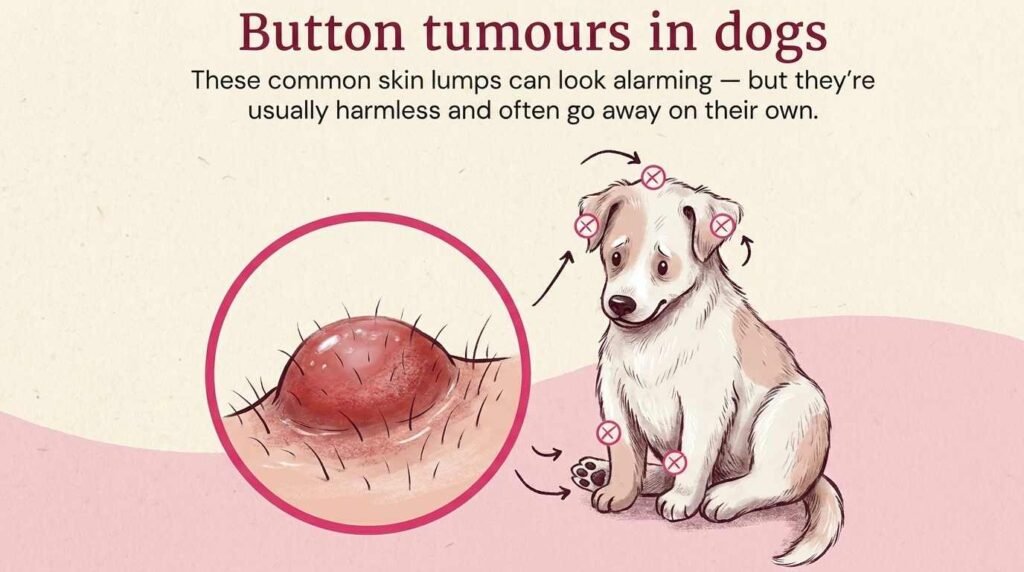
They appear as raised, pink or red, flashy-looking domes that show rapid growth and may then ulcerate before regressing. These are most common in dogs aged 1 to 2 years and are very rare in senior dogs.
These usually appear on the head, ears, and legs. In 80 to 90% of the cases, they regress spontaneously within 2 to 3 months as the immune system recognises them as abnormal cells and destroys them. Generally, no treatment is required; they can be removed surgically if they persist.
8. Papillomas (Warts)
Papillomas are usually rough, cauliflower-like growths that most commonly occur around the mouth, eyes, and footpads, with canine papillomavirus as the primary causative agent. They are most common in puppies and young dogs and resolve easily within 1 to 5 months as the immune system develops, but they can interfere with eating and lead to secondary infection.
9. Perianal Adenomas Appearance
These are firm masses that appear near the anus in a lobulated form. There are testosterone-dependent characteristics, mostly exclusive to male dogs, that can be reversed by castration. In addition, the residual mass can be surgically removed.
10. Mast Cell Tumours (MCTs) — MOST COMMON MALIGNANT SKIN TUMOR IN DOGS
Mast cell tumours contain histamine and heparin, which can cause local inflammation, gastric ulcers, and anaphylactic shock, and can also affect blood pressure and clotting when released.
In appearance, they can be hairless raised bumps, subcutaneous masses, or normal skin that is red, inflamed, and dilated, ranging in size from 1 cm to 10 cm or more. The breeds at greatest risk are Boxers, Boston Terriers, Golden Retrievers, Labrador Retrievers, Shar-Peis, Pugs, and Staffordshire Bull Terriers.
| Feature | Grade I (Low) | Grade II (Intermediate) | Grade III (High) |
|---|---|---|---|
| Cellularity | Low | Moderate | High |
| Mitotic figures | Rare (<1 per HPF) | Variable | Frequent (≥7 per HPF) |
| Invasiveness | Well-demarcated | Variable | Poorly demarcated, invasive |
| Metastasis risk | <5% | 15-30% | 55-96% |
| Median survival (with surgery) | >1500 days (most cured) | 300-800 days | 80-180 days |
| 1-year survival | 93% | 60% | 16% |
16. Soft Tissue Sarcomas (STS)
STS could be of multiple types, including fibro, peripheral nerve sheath tumour, hemangiopericytoma, and malignant fibrous histiocytoma. They can appear as firm, fixed lumps in underlying tissues, while others feel like a pebble under the carpet.
STS are locally invasive, with roots extending beyond palpable margins. There could be high chances of recurrence if not completely removed due to metastasis and eventually affect the lungs in 15 to 25% cases. The only treatment is surgical removal or radiation therapy; however, chemotherapy is still controversial.
17. Squamous Cell Carcinoma (SCC)
SCC appears as irregular, crusty, ulcerated plaques or in modular form and could lead to non-healing wounds. The risk factors are sunburn, thin-furred areas, papillomavirus, or any chronic inflammation. They usually appear on the nose, nail beds, abdomen, scrotum, or vulva.
The only treatment is surgery; however, radiation or chemotherapy can also be performed in advanced cases.
18. Melanomas
These appear as black or dark brown pigmented masses, but may also be described as pigmented in some cases. They are most common in dogs as oral melanomas.
They show very aggressive metastasis in lymph nodes and lungs, and have a poor prognosis. In dental melanoma, they are often benign. The treatment protocol includes surgical removal, vaccine or radiation therapy. Despite aggressive. Therapy shows a guarded prognosis.
Treatment
The treatment of a tumour depends upon its type. Once the veterinarian has diagnosed the type of lump found under the dog’s skin, it is easy to treat with the appropriate procedure. Based on the data presented above, I have categorised the treatment protocol into four categories. Here are the details.
1. Protocol A: Suspected Benign Mass
In this case, the conservative management pathway is followed. First step is confirmation that it is an adipose cell, no atypia. Photograph, measure, and record the location of the lump. Make a schedule to recheck it every six months. Initially, if it is stable, then an annual check is done. Also, educate the owners to continuously check for any new symptoms or rapid growth. Follow the surgical method only if there are suspicious changes or an increase in size.
2. Protocol B: Low-Grade Mast Cell Tumour
First of all, aspirate the tumour with a long needle, do the basic bloodwork, and perform an ultrasound if necessary. If it is confirmed to be a mast cell tumour, perform surgery. To check whether it has been excised completely, perform histopathological confirmation, then monitor it for 6 months to 2 years.
If clean margins are achieved, the cure rate exceeds 90%. If the margin still contains numerous cells, options include a second surgery, radiation therapy, or close monitoring (considered controversial).
For medication, consult the veterinarian. He can prescribe any antihistamine or gastroprotectants to reduce the risk of granulation. It has an excellent prognosis, and most of the dogs are cured with surgery alone.
3. Protocol C: Intermediate-Grade Mast Cell Tumour
In this case, due to variable behaviour, treatment intensity is individualised based on the following,
- Mitotic index (≤5 vs. >5 per 10 HPF)
- c-KIT mutation status
- Location (visceral vs. integumentary)
- Surgical margin status
- Client goals and financial constraints
The first-line treatment is surgical excision with wide margins. However, other approaches can also be considered. These are as follows,
- Radiation therapy (if margins are incomplete or the location is unresectable)
- Definitive: 42-48 Gy in 15-21 fractions (cure intent)
- Palliative: 8 Gy weekly × 4 treatments (symptom control)
- Chemotherapy (controversial benefit; clearer role for gross metastatic disease)
- Vinblastine + Prednisone (most common protocol)
- Lomustine (CCNU) — especially for visceral/nodal disease
- Newer combination trials ongoing
- Tyrosine Kinase Inhibitors (TKIs)
- Toceranib phosphate (Palladia®) — FDA-approved; first-line targeted therapy
- Masitinib (Kinavet®) — Available in some regions
- Particularly indicated for c-KIT mutant tumours or when chemotherapy is contraindicated
- Cost: $200-$500/month ongoing
- STELFONTA® (tigilanol tiglate) — Intratumoral injection causing tumour necrosis
- Ideal for: Unresectable locations, nonsurgical candidates, owners declining surgery
- 75-87% complete response in clinical trials
- Single or double injection protocol
- Side effect: Significant local inflammation/necrosis during healing (manageable)
4. Protocol D: High-Grade Mast Cell Tumor
In this condition here is the complete recommended approach for treatment.
- Complete staging (mandatory)
- Surgery
- Chemotherapy (strong recommendation)
- Vinblastine/Prednisone OR Lomustine-based protocols
- Consider novel combination trials if available
- TKI therapy — often combined with chemo or sequenced
- Radiation for local control if primary not completely resectable
- Supportive care: Gastric protectants (long-term), antihistamines PRN, pain management
- Palliative/hospice focus when appropriate: Prioritize comfort over further anti-cancer therapy
Realistic Expectations (based on 2025 data):
- 6-month survival: ~69%
- 1-year survival: ~50%
- 2-year survival: ~30%
- Median overall survival: 317 days (range 20-3,041 days reported)
- 75% of dogs will experience disease progression despite aggressive therapy
Decision Flowchart
📊 Quick Assessment Algorithm
Breeds At Most Risk
There are certain breeds of dogs that are that more risk of developing lumps under the skin as compared to other breeds here is the detail of those breeds that are at most risk.
| Breed | Elevated Risk For | Recommended Screening |
|---|---|---|
| Boxer, Boston Terrier, Pug, Shar-Pei | Mast cell tumors | Monthly checks; FNA any new lump immediately |
| Golden Retriever, Labrador Retriever | MCTs + Hemangiosarcoma | Monthly checks; annual ultrasound after age 7 |
| Labrador, Doberman, Sheltie | Lipomas (benign but monitor for changes) | Quarterly checks; document all masses |
| German Shepherd, Golden Retriever | Hemangiopericytoma / STS | Monthly checks; low threshold for biopsy of firm fixed masses |
| Cocker Spaniel, Poodle, Miniature Schnauzer | Sebaceous gland tumors | Watch for wart-like growths; biopsy if changing |
| Weimaraner, Bernese Mountain Dog | Histiocytic diseases | Know breed-specific presentations; prompt biopsy of skin masses |
| Dalmatians, Beagles, Whippets, Bull Terriers | Squamous cell carcinoma (sun-related) | Sun protection; early biopsy of non-healing sores |
Conclusion
There are some strategies that you can follow to prevent any of such circumstances. These include proper neutring of your dog at proper time healthy weight management and avoid sunburns.
I have tried to discuss almost all the conditions and their solution solutions in this blog post. If you still have any questions, do let me know in the comment section thank you.